Leg Bones Diagram / 16 best Bones in the Leg images on Pinterest | Human body ... / You'll learn about the muscles, bones, and other structures of each area of the leg.
Leg Bones Diagram / 16 best Bones in the Leg images on Pinterest | Human body ... / You'll learn about the muscles, bones, and other structures of each area of the leg.. Click now to learn more about the bones, muscles, and soft tissues tibia: The patella in the knee; Time to jump right into the biggest and strongest bones in the human body. Here are a few anatomical plates about the leg and the foot. The foot bones shown in this diagram are the talus, navicular, cuneiform, cuboid, metatarsals.
Its lower end helps create the knee joint. The patella in the knee; The foot bones shown in this diagram are the talus, navicular, cuneiform, cuboid, metatarsals and calcaneus. At the microscopic level, this hard outer. This page is about leg bones diagram,contains aluminium plant safety:
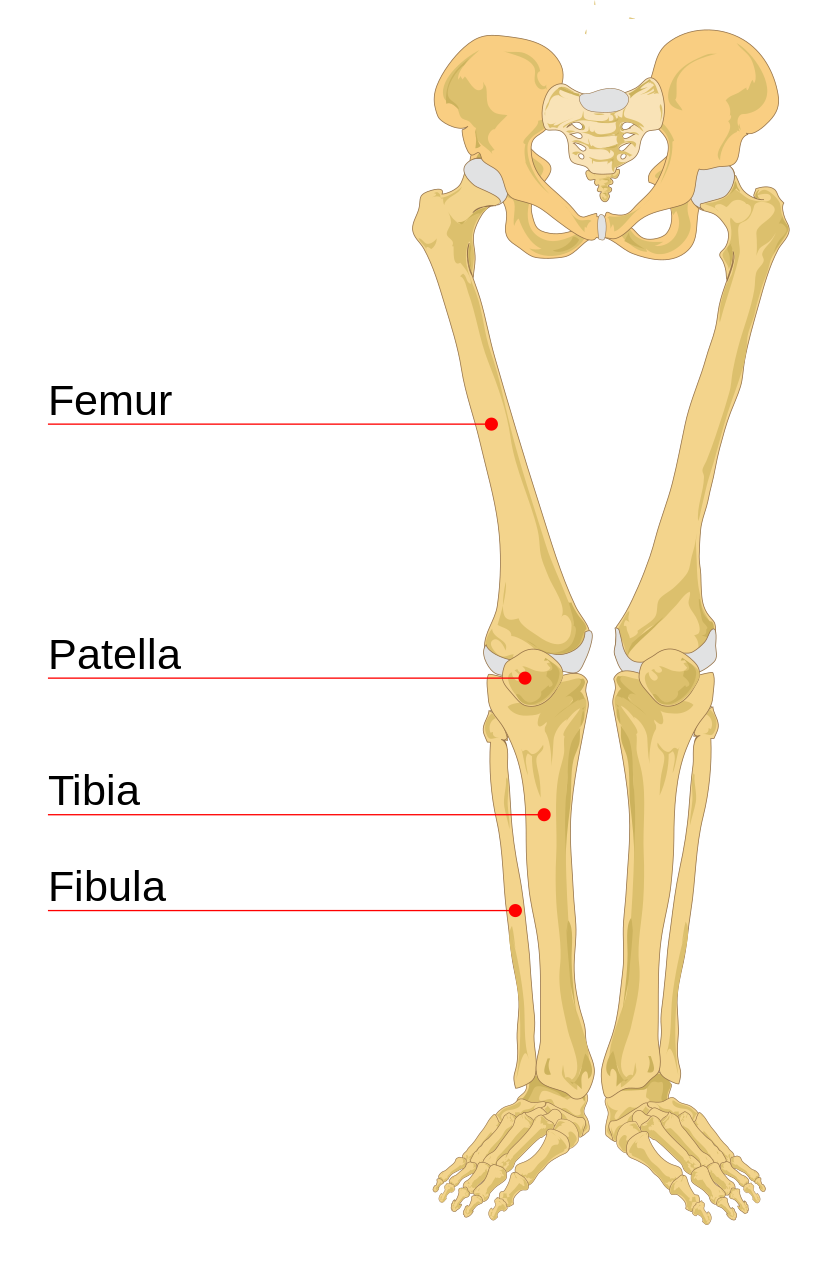
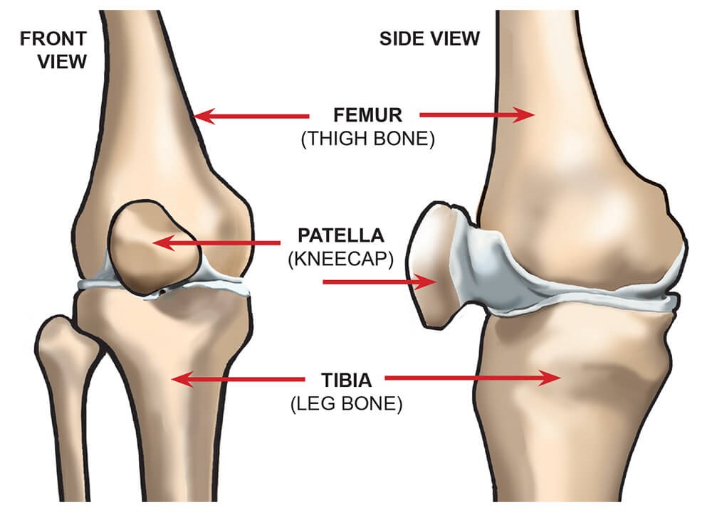
File:Human leg bones labeled.svg - Wikipedia from upload.wikimedia.org Each leg is made up of four bones. The femur in the thigh; At the same time, the bones and joints of the leg and foot must be strong enough to support the body's weight while remaining flexible enough for movement and balance. Use the leg bones diagrams to learn the names of the leg bones. Most bones (particularly the long bones of the arms and legs — which make up the appendicular skeleton) have a hard outer shell known as cortical bone. Your leg bones are very large and strong to help support the weight of your body. At the microscopic level, this hard outer. Visit kenhub for more skeletal system quizzes.
Normal leg bones are relatively straight, but those affected by paget's disease are porous and figure 9.
At the microscopic level, this hard outer. Your legs are two of your most important body parts. Its lower end helps create the knee joint. The bones of the leg are the femur, tibia, fibula and patella. These simple labelled diagrams of the bones of the lower legs and feet and the bones of the arms and hands this diagram shows the skeletal structure of the leg (anterior view) and foot (dorsal view). The femur in the thigh; Nervsystemet anatomy, diagram & function | health. Your leg bones are the longest and strongest bones in your body. The foot bones shown in this diagram are the talus, navicular, cuneiform, cuboid, metatarsals and calcaneus. When you stand or walk, all the weight of your upper body rests on them. This page is about leg bones diagram,contains aluminium plant safety: The foot bones shown in this diagram are the talus, navicular, cuneiform, cuboid. Click now to learn more about the bones, muscles, and soft tissues tibia:
Most relevant best selling latest uploads. The human leg consists of 8 bones, 4 per leg. Its lower end helps create the knee joint. Learn how to draw the femur, patella, tibia, and fibula in this lesson! They allow you to move and provide support for your upper body.
The Art of Matt Gutpell: Bones Finally Done for Life ... from photos1.blogger.com Learn how to draw the femur, patella, tibia, and fibula in this lesson! Ankle and foot pain massage therapy connections. The foot bones shown in this diagram are the talus, navicular, cuneiform, cuboid, metatarsals and calcaneus. This page is about leg bones diagram,contains aluminium plant safety: Visit kenhub for more skeletal system quizzes. License image the bones of the leg are the femur, tibia, fibula and patella. They allow you to move and provide support for your upper body. Diagram of blood and nerve supply to bone.
Your leg bones are the longest and strongest bones in your body.
Blood vessels and nerves enter the bone. Master leg and knee anatomy using our topic page. Diagram of blood and nerve supply to bone. Use the leg bones diagrams to learn the names of the leg bones. Ankle and foot pain massage therapy connections. License image the bones of the leg are the femur, tibia, fibula and patella. The human leg consists of 8 bones, 4 per leg. Your legs are two of your most important body parts. Click now to learn more about the bones, muscles, and soft tissues tibia: He leg's main function in the human is for locomotion and support of the rest of the body. This page is about leg bones diagram,contains aluminium plant safety: Time to jump right into the biggest and strongest bones in the human body. At the same time, the bones and joints of the leg and foot must be strong enough to support the body's weight while remaining flexible enough for movement and balance.
Blood vessels and nerves enter the bone. The patella in the knee; They allow you to move and provide support for your upper body. When you stand or walk, all the weight of your upper body rests on them. Master leg and knee anatomy using our topic page.
Knee joint - is it really something to fear? | UKRunChat from www.ukrunchat.co.uk The foot bones shown in this diagram are the talus, navicular, cuneiform, cuboid. Human anatomy diagrams show internal. Click now to learn more about the bones, muscles, and soft tissues tibia: However, the definition in human anatomy refers only to the section of the lower limb extending from the knee to. Visit kenhub for more skeletal system quizzes. They allow you to move and provide support for your upper body. The human leg consists of 8 bones, 4 per leg. The human leg, in the general word sense, is the entire lower limb of the human body, including the foot, thigh and even the hip or gluteal region.
However, the definition in human anatomy refers only to the section of the lower limb extending from the knee to.
Each leg is made up of four bones. The bones of the leg are the femur, tibia, fibula and patella. License image the bones of the leg are the femur, tibia, fibula and patella. The femur in the thigh; Human anatomy diagrams show internal. Click on the figures for a detailed view you will find the pelvic bones in the hip; Normal leg bones are relatively straight, but those affected by paget's disease are porous and figure 9. However, the definition in human anatomy refers only to the section of the lower limb extending from the knee to. The largest and most medial leg bone, forming both the knee and ankle joints. Your leg bones are the longest and strongest bones in your body. Most bones (particularly the long bones of the arms and legs — which make up the appendicular skeleton) have a hard outer shell known as cortical bone. The foot bones shown in this diagram are the talus, navicular, cuneiform, cuboid. Click now to learn more about the bones, muscles, and soft tissues tibia:


No comments:
Post a Comment